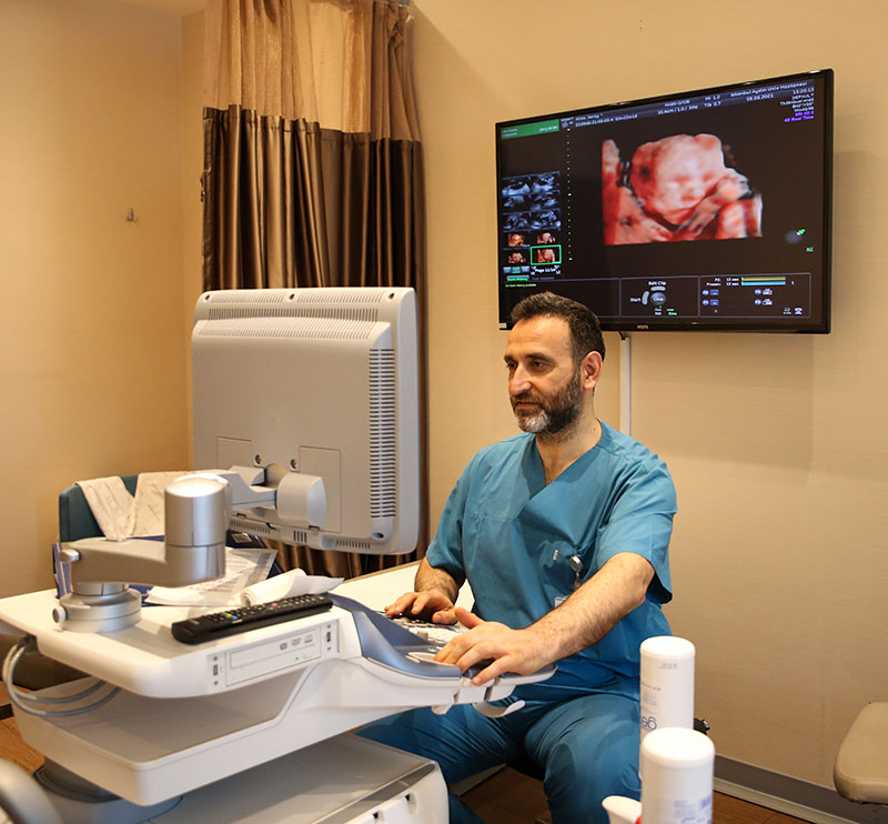
While black and white imaging (B-mode) used in daily ultrasound examinations provides a 2D image of the fetus, modern ultrasound technology can produce lifelike 3D or 4D motion images of the fetus.
3D/4D examination sections are not an essential requirement for a detailed ultrasound examination, but they do make it easier for parents to understand some anomalies, especially facial pathologies/diseases.
Not only can 3D/4D technology be used to show the baby's face, but depending on the different technological modalities of the ultrasound machine, 3D/4D technology can also help assess tissues such as the heart, blood vessels, bones, and brain tissue.
With 3D/4D imaging of the fetus, the baby's face and movements can be seen from 20 weeks of pregnancy. Optimal assessment is between 25 and 32 weeks of gestation, after which imaging success decreases due to the baby's growth and relative fluid loss.
Since all imaging ultrasound systems (2D, 3D and 4D) are based on sound waves, no harm to the mother or baby is expected.


