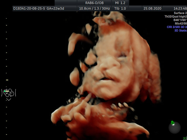
Although a detailed ultrasound examination is performed between 18 and 23 weeks of gestation, it is preferably performed between 20 and 23 weeks. The most important reason for performing this examination at these weeks is that the anatomy of the fetus (baby in the womb) can be examined most clearly at these weeks. In earlier or later weeks of pregnancy, the examination may be inadequate. However, if an anomaly is suspected, a detailed examination should be performed regardless of the week of pregnancy. A detailed ultrasound examination of the fetus is a procedure that should be routinely performed on every expectant mother, preferably as much as possible.
This screening examination differs from pregnancy ultrasound, which assesses fetal development during routine pregnancy examinations. A detailed ultrasound examination is performed by perinatologists between the 20th and 23rd weeks of gestation to detect structural anomalies of the fetus. The incidence of major congenital structural anomalies in infants is 2-3%. A thorough ultrasound examination of the fetus can rule out 80% of these anomalies. However, this rate may vary depending on the quality of the ultrasound image, the patient's adipose tissue, the position of the fetus, and the week of gestation. The examination time must be sufficiently long (at least 30 minutes) to allow a detailed examination of each organ. The main goal is to provide the best possible postnatal care for the child if the pregnancy continues, as well as to provide suggestions and instructions according to the possible anomalies that may be detected after the examination. Even the treatment of some diseases in the womb is planned in this way.
The detection rate of minor anomalies, which are more common in the population, are small, and do not affect organ function, is lower: these include some finger anomalies, small heart holes, isolated cleft palate, genital deformities, nostril obstruction, and absence of an anus. Diseases that do not cause anatomical abnormalities in organs and systems or are associated with mild dysfunction may not be detected on ultrasound examination.
Cleft Lip

Although one of the main purposes of detailed ultrasonography is to detect genetic diseases associated with mental retardation, some genetic syndromes associated with mental retardation may not show findings on ultrasound. In addition, there may be some pathologies/diseases that are not visible during the week of detailed ultrasound examination or may be detected only in later weeks (e.g., brain hemorrhages, skeletal dysplasia, tumors, etc.).
This is a combined examination that includes fetal echocardiography to assess the function and structure of the heart, Doppler sonography to assess the function of the placenta and uterine vessels coming from the mother, and cervical measurement to rule out possible cervical insufficiency.
However, a detailed ultrasound examination of the fetus is incorrectly referred to as a “color ultrasound,” and it should not be mistakenly assumed that the images obtained will be in color. The images from this examination are similar to the images obtained by the gynecologist during routine examinations. The difference is that these examinations are performed by experienced specialists using equipment of high image quality. In addition, the baby’s face and other organs can be examined using 3- and 4-dimensional ultrasound techniques.


